|
How does Bethlem Myopathy make Julie different? What things about Julie are the same?
|
 |
|
Really, the question is how does everyone's body know how to make itself? The answer is DNA (deoxyribonucleic
acid)--DNA is the "instruction manual" for all life. In people, DNA is over 99% the same in everyone. It's the
little differences in that 1% that make us different from each other.
|
| Taken from: http://www.koshlandsciencemuseum.org/exhibitdna/intro02.jsp |
There is a section of DNA in people with Bethlem Myopathy that tells the body to make a connective tissue called "Collagen
VI" wrong. This section is called a "gene." Because Collagen VI is made wrong, it affects how the muscles grow
and work in people with Bethlem Myopathy. Scientists and doctors are still trying to understand just how this problem
with Collagen VI affects the muscles so maybe they can find a way to treat the problem or cure it. That's still a long
way off, though.
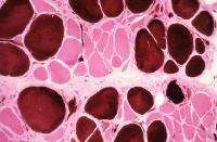
What we do know is that the muscles of people with Bethlem Myopathy look different under the microscope. Here is
a picture of normal muscle and then a picture of muscle from a person with a myopathy (not from Julie--these pictures are
from science articles).
|
| Taken from: http://jmg.bmjjournals.com/cgi/content/full/42/9/673 |
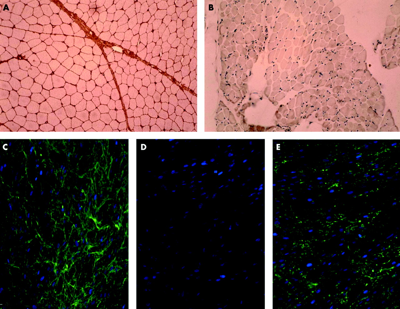
These pictures are from a person with normal muscles, a person with a more severe form of Bethlem Myopathy called Ullrich
Muscular Dystrophy, and from a person with Bethlem Myopathy. (Again, none of these pictures is from Julie, rather from
a science article.)
The top two pictures are of muscle, on left, from a person with normal muscles and on the right, from a person with Ullrich
Muscular Dystrophy. You can see on the left that the muscle cells are nice and even, approximately the same size, and
have lots of Collagen VI in between the muscle fibers--the Collagen VI is stained red so you can see it. On the right,
there is almost no Collagen VI and the muscle fibers are different sizes and not as nicely organized. In a person with
Bethlem Myopathy, it would look something like the one on right, but with a bit more Collagen VI in the spaces.
The bottom pictures are from skin cells and show how Collagen VI should look on the left. The Collagen VI is stained
green and looks plentiful and makes a kind of web, connecting everything together (hence the word, connective tissue).
The middle picture is from a person with Ullrich Muscular Dystrophy and you cannot see any Collagen VI. The picture
on the right is from a person with Bethlem Myopathy. You can see some green-stained Collagen VI, but there isn't much
of it and it seems like it is just hanging out there in little bunches rather than forming that nice web that it should form.
In person with Ullrich Muscular Dystrophy, Collagen VI doesn't form at all. The gene responsible puts a different
protein in the wrong place and that screws up the formation of Collagen VI. In a person with Bethlem Myopathy, Collagen
VI does form, but not as well as it should. The gene responsible puts a different protein in the wrong place so that
some Collagen VI forms, but it doesn't look right and doesn't do its job (whatever that is!) as well as it should.
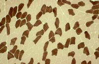
Another thing that is affected is how the muscles form:
|
| Normal muscle--type 1fibers are tan and type 2 are brown |
|
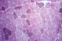
| Myopathic muscle--many more type 1 fibers (light color) than type 2 (dark color) |
|
| Here, the type 1 and type 2 fibers are of different sizes. They should be all the same size. |
http://www.emedicine.com/pmr/topic65.htm
and show typically myopathic findings on muscle biopsies. The top picture is of normal muscle. There is a pretty
even amount of the type 1 and type 2 muscle fibers and the fibers are pretty much the same size. The slide below represents
a finding similar to Julie's muscle biopsy--there are more type 1 fibers (light color) than type 2 fibers (dark color).
The next slide shows another finding that was similar to Julie's muscle biopsy--the size of the muscle fibers varies greatly.
Some are much smaller than the others. They should all be close to the same size like in the top slide.
|
|
|
|
|
|
|
|
|
|
|
|
|
| Julie and her pups, Kiwi and Cocoa |
So how does Bethlem Myopathy make Julie the same as
other kids and how does it make her different?
In most ways, Julie is the same. She has fingers and toes, thoughts and feelings, likes
and dislikes, and pretty much everything that any other nine year old has. Everyone is different is some ways,
like the color of their hair and which foods like they to eat. Julie is no exception!
In a few ways, Julie is different. Mainly, Julie is different in how her muscles work.
Because her muscles look different under the microscope, like in the pictures on this page, Julie is not as strong or as fast
as other kids her age. Her mind knows what she wants her muscles to do, but her muscles take longer to learn to do tricky
things, like dribbling a basketball and running fast.
Her muscles are not as strong as other kids, either. Most of the time they are strong
enough for what Julie wants and needs to do every day, like walk around school, open and close her desk, pick up a book to
read, etc. Sometimes her muscles are not strong enough and then Julie needs help, like with opening a lid or a really
heavy door. Julie's muscles in her torso are most affected by her myopathy and that means that it can be hard for Julie
to keep her balance. This is why it is so upsetting to Julie when she gets bumped--it feels like she might fall down
and that's a scary feeling.
Another way Julie is different is that she is very flexible! Her joints can open farther
and bend back farther than most people's joints can. One joint that is not as flexible, however, is her knees.
The muscle behind her knees, called the hamstring, seems to want to tighten up and make it hard for Julie to straighten her
knees all the way. This happens to people with Bethlem Myopathy and, at times, we do stretches with Julie every
night to keep her knees flexible. So far, it's working!
Finally, Julie's muscles get tired more quickly than other kids. It's not the same kind
of tired like when you need to sleep. It's the kind of tired that you feel when you have been running a long way and
your muscles won't let you run anymore and you have to stop and walk or rest. Because Julie's muscle are different,
they have to work harder to do things and they use up Julie's energy more quickly. This is why it's important that Julie
has her chairs at school that support her body so it doesn't have to work as hard and so she won't get as tired as quickly.
This is also why it's important that Julie gets to rest her body when she needs to.
The most important way in which Julie is just like everyone else is that she has feelings.
She is not a robot! She can feel happy, sad, mad, proud, etc. She is a great kid and a good friend.
This page was created to explain to Julie's class about Bethlem Myopathy and how it makes Julie different.
It is not intended as medical advice and I make no promises about the accuracy of the information! It represents my
understanding and interpretation of the information and the manner in which I decided to simplify it for an audience of fourth
graders.
|