|
In August of 2002, we were faced with two different opinions from highly regarded experts at Children's Hospital of Philadelphia.
A geneticist was "certain" that Julie has Ehlers-Danlos Syndrome (EDS), a connective tissue disorder, causing her
hypermobility and hypotonia. Julie's neurologist, a neuromuscular expert, was "more certain than ever" that Julie
has some kind of congenital myopathy. The only way to definitively diagnose Julie would be with a muscle biopsy.
My husband Mark and I pondered this decision for about three weeks. Foremost in mind was the future, especially the near
future of elementary school. The geneticist had cautioned us to protect Julie's joints as she grew up by limiting high impact
sports and other activities, such as the monkey bars on the playground, that might overstress her joints. However, the neurologist
placed no such restrictions on Julie's future in the case of a congenital myopathy. He felt that she should always be encouraged
to do as much as she can do, given her motor delays and fatigue. Presenting Julie's needs to our public elementary school
was also on our minds. It did not seem to us that going into the school and saying "well, she might have EDS, she might
have a myopathy...we're not sure but she will need these accomodations" would be the most prudent course of action.
We felt that planning for Julie's education accomodations would be easier with a definitive diagnosis to help guide us and
her educational team.
Mark, as a physician, was concerned about how specific the muscle biopsy would be as a medical test. We learned that
three results were possible. One, it could certainly come back showing nothing but completely normal muscle. However, both
we and the neurologist did not think that was likely at this point and we would not have gone ahead with a biopsy if it was
likely to come back completely normal. Second, it might come back with a definitive diagnosis of one of the documented congenital
myopathies or possibly, as the neuro put it, "a surprise" of some kind of neuromuscular condition that had not been
previously suspected in Julie's case. Third, there was a chance of a non-specific result. That is to say, that the biopsy
would show some kind of abnormality that was not specific to any documented diagnosis.
The final factor that helped us make our decision to go ahead with the biopsy was my own level of anxiety over Julie having
a diagnosis. For the three years since her hypotonia was recognized by the pediatrician, we both had been comfortable not
knowing the cause for Julie's hypotonia because she always made progress and having a diagnosis would ultimately not change
anything we would do for her. Now, with the possible EDS diagnosis, there were things that we would do at least somewhat
differently for Julie if we needed to protect her joints from EDS. But most of all, I was starting just to NEED to know.
In my heart and in my gut, I needed to know what was causing her problems so I could go on with the future and help her as
her mother in the most informed way possible. I also wanted to be able to explain her differences to her once she started
asking about them and I wanted to be able to tell her with as few "I don't knows" as possible.
So the decision was finally made and I contacted the neurologist to find out how to proceed. He put us in touch with
a general surgeon who does 95% of the muscle biopsies for the neurology department. I have since learned that it is important
to have a surgeon who has done many biopsies. When all is said and done, it is very minor surgery, but it is still quite
specialized and certainly something you want done right the first time! We also agreed to have a skin biopsy done at the
same time to be evaluated in a CHOP research lab where research is being conducted into the relationship between problems
with collagen 6 and neuromuscular problems.
The surgery consultation went well. The surgeon was very patient with us (ok, me!) and answered all our anxious questions.
We were told that the skin biopsy would be taken at the incision site for the muscle biopsy, so there would be only one scar.
The surgeon, in consultation with the neuro, chose to do the biopsy in Julie's right quadrocep because the PT at the neuro's
office had noticed that Julie's right side was somewhat weaker than the left and therefore possibly more likely to show results.
Julie's weakness, at this age, seemed pretty uniform, so the surgeon and the neuro were confident that the biopsy site would
be appropriate.
We also discussed Julie's anesthesia. From Dr. Charlotte Thompson's book, "Raising a Child With a Neuromuscular
Disorder", we were already aware that a child with an undiagnosed myopathy was at risk for a complication of anesthesia
called malignant hyperthermia. It seems that the most widely used inhaled anesthesia, Halothane, and its related class of
anesthesias, can cause a potentially fatal reaction within the body where the body's temperature rises very quickly, damaging
internal organs. It is possible to have the gene for malignant hyperthermia and not have any kind of myopathy, but people
with Central Core Myopathy and some with Minicore (aka Multicore) Myopathy are at increased risk for malignant hyperthermia
because the gene for these conditions is near the malignant hyperthermia gene. So, since we did not know which myopathy Julie
might have, we were assured that IV anesthesia would be used and that the risk of problems was virtually none. We were also
told that Julie would be intubated (a tube would be put in her throat to help her breathe) while she was under anesthesia.
In all, the biopsy was expected to take about a half an hour from first cut to finish.
The final thing we discussed with the surgeon is how to manage Julie emotionally through the process. He gave us a booklet
published by the hosptial for day surgery patients and suggested that we read it with her. He also said not to even call
it surgery or an operation and to say that she was having a "muscle test". He had a four year old daughter and
said that is what he would tell her.
We finished our surgical consultation and were surprised to find that we could schedule Julie's biopsy for a week later!
I was glad that it would be over soon, but my anxiety went through the roof. It was a long week full of trouble sleeping
and stomach aches. My mother agreed to come in to care for my older daughter Robin the day of the surgery which was a tremendous
help.
The day of the surgery, we had to be at CHOP at 6am (ugh). I did not sleep well the night before, but somehow we did
manage to get up and get out to arrive more or less on time. We picked a sleeping Julie up from her bed at 5:30am and put
her in the car, hoping she would go back to sleep. She did not and chatted with us the whole way to the hospital! She was
not allowed to have anything to eat or drink and, luckily, she did not ask for anything. Of course, the first thing you do
is hurry up and wait! We were brought back to the pre-op room and Julie was given the option of her nightgown or hospital
pajamas. She chose her nightgown and they weighed her and took her temperature and blood pressure. We brought along some
toys, her precious Blankie, and a favorite stuffed dog of hers named BJ. After that, we had about a forty minute wait until
the anesthesiologist came in and gave us the run down on how things would go. He told us that Julie would get some "giggle
medicine", aka Versed, and would get tired and loopy and relaxed before they took her back to surgery. Once in the OR,
they would give her a little nitrus oxide and then once she was asleep, they would give her an IV and intubate. Once the
biopsy was over, one of us would be allowed back to the post-op room to be there once she woke up.
|
 |
|
|
|
|
|
Julie got the Versed and a little sip of apple juice to wash it down at about 7:30am. She sat in my lap playing with BJ and
over about fifteen minutes, I noticed her getting heavy and limp. She could still control her muscles, though, and was having
BJ sniff around and bark while we cuddled. Finally, they felt she was feeling the full effect of the Versed and the brought
a guerney to take her to surgery. Julie let us place her, Blankie, and BJ on the guerney without any anxiety and as they
rolled her away, we could hear her barking all the way down the hall!
We were then shown the parents' waiting room where we sat and talked and tried to pass the time. Mark was great--he prattled
on and on about his work and the crazy office politics he was dealing with to distract me from the my anxiety. After about
twenty minutes, a nurse came out and assured us that Julie had gone under anesthesia with no problems, barking the whole time!
That's my Julie! She said that the biopsy was starting and should take about a half an hour from start to finish. It was
nearly an hour, though before the nurse came back and brought me to see Julie.
Luckily, one of my myopathy email list friends had told me about "emergence delirium" from anesthesia. It seems
that small children can become very upset and disoriented for 15 to up to 90 minutes after anesthesia. So when I heard Julie
screaming from down the hall, I at least knew to expect it! I found Julie red eyed and screaming very angrily, harnessed
to her bed so she wouldn't fall or more likely jump off, wearing hospital pajamas and with an IV board taped to her left wrist.
She was furious about the IV board and was waving her hand around and banging it into the bed, the railing, and my head (!)
trying to get it off. She was also mad that she was no longer wearing her nightgown. However, for the first fifteen minutes
it was clear that she was not fully aware of what was going on around her. She seemed to answer me as I tried to console
her, but she did not meet my eyes (or anyone's) and continued to flail. After about 15-20 minutes, she started to become
more aware and in ten minutes more, calmed down enough to move to the recovery room.
In the recovery room, Julie was calm and alert and let us read to her for a while. She asked for juice, at last, and
then had two boxes of cereal. We brought her some new Dora the Explorer stamps and she made some pictures and played with
some toys while the nurses monitored her for any post surgical or anesthesia problems. Julie did really well and only complained
that her leg hurt while we were in recovery. They gave her a dose of tylenol with coedine and she was on her feet, walking
to her stroller when it was time to leave. We went home by way of CVS for more coedine and Julie fell asleep in front of
the tv for most of the afternoon.
We were told by the anesthesiologist that Julie should stay home from school and her activities the day after just to
make sure she was fully recovered from the anesthesia. But two days later, Julie went back to school, OT and ballet with
no problems. The surgeon did say to try to limit the amount of jumping and tumbling around that she does for about a month
to allow the biopsy site to heal, but other than that, to let her do her normal routine.
|
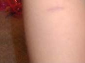
| Julie's muscle biopsy scar on her upper right thigh. |
We gave Julie the tylenol with codine for the rest of the day of her surgery and the day after, although she did not complain
of pain after we left the hospital. After she went back to school, we waited to see if she complained of pain and she did
not so no more medicine was necessary. The surgeon said that it would not be that painful and that it would feel like a deep
bruise for a few days, if anything. Julie proudly went around lifting up her dress and showing everyone the butterfly bandage
covering her "muscle test"!
Then, the wait for results began. The neuro told us that the muscle biopsy results would take about four weeks and the
skin biopsy results would take at least eight weeks. We called after four weeks and were told that the initial muscle biopsy
results were "mildly abnormal and non-specific" (yes, the dreaded non-specific result!). They did not find any
signs of a dystrophy and there were no signs of any kind of storage problems in the muscle (no fat, no glycogen). There were
no atrophic or hypertrophic fibers seen, nor did the microscope find any rods, cores, or other signs of myopathy. The one
thing they did find was a disproportion in size between some of the type 1 and type 2 muscle fibers, mixed in with fibers
of the expected size. This finding, which in the UK might be called "CFTD--congenital fiber type disproportion",
was what the neuro considered mildly abnormal and non-specific. The neuro said that the skin biopsy results would take at
least another 4-6 weeks because the research lab was growing "fibroblast cultures" from the skin cells to study
and that takes quite a long time.
With help from my myopathy friends and some email from a specialist in California, I later pressed the neuro about doing
electron microscope (EM) studies of the muscle biopsy. He told me initially that they had not been done because it would
be like looking for a needle in a hay stack. However, the specialist from California insisted that there are things that
could show up on EM study that would not show up under the regular microscope. The neuro agreed that the studies should be
done but was waiting for the skin biopsy results before proceeding.
The last I have heard as of the end of February, 2003, is that the EM study that the neuro thought had been done was in
fact not done until this month. I have been consulting with a neuromuscular expert in California for a second opinion and
our hospital finally sent her Julie's muscle biopsy slides and EM pictures. She was not happy with the quality of the slides
and we may consider having them redone by her friend, the muscle biopsy world expert, in the UK. The CA doc thought she might
have seen central cores or minicores on the EM pictures, but she can't tell if it is a real finding or due to inadequate preparation
of the sample. Her UK friend will be able to take a look in a few weeks. The skin biopsy is still being evaluated in the
research lab at CHOP. Nothing had happened on the skin biopsy evaluation until February also. Regardless of how these results
are trickling in, the neurologist feels that Julie's clinical picture along with what we do have from the muscle biopsy is
strongly suggestive of myopathy, so for now he has dubbed her diagnosis "congenital myopathy, nos" (nos means not
otherwise specified). The main diagnosis that the neuro suspects is Bethlem Myopathy which is quite rare, but nevertheless
a possibility given Julie's hypotonia, hyperflexibility, and congenital torticollis. Apparently it is the skin biopsy results
that are instrumental in diagnosing Bethlem Myopathy.
The waiting for results has been tedious, but not nearly as bad as the month it took to make the decision to go through
with the biopsy and then to actually go through with it. I am frustrated, though, because I am starting to suspect that Julie's
condition may be scraping against the ceiling of what is known to medical science at this point. I do not regret doing the
biopsy, but I am worried that we will end up with a "mildly abnormal, non-specific" result. I am also not yet clear
on whether any of this rules in or out EDS.
June, 2003...the diagnosis is in! Click on "The Diagnosis" to read more!
|
|
|
|
|